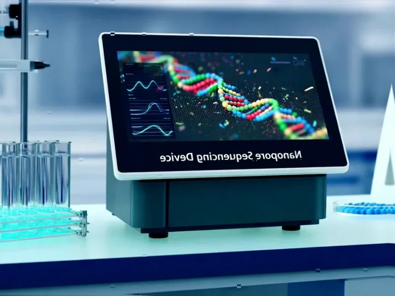
According to Phys.org, researchers from Bigelow Laboratory for Ocean Sciences and Atrandi Biosciences have developed a breakthrough method called environmental microcompartment genomics that vastly improves single-cell genetic sequencing. In their Nature Microbiology study, they sequenced over 2,000 particles from just 300 nanoliters of seawater from the Gulf of Maine, increasing throughput by an order of magnitude compared to traditional methods that process only 384 particles per run. Lead author Alaina Weinheimer said the approach increases both quantity and quality of data while reducing costs, and early testing shows it works on sediment and soil samples too. The method captured viruses of all sizes simultaneously, including the Naomiviridae family that other approaches would have missed entirely.


The tiny bubble revolution
Here’s how this actually works – and it’s pretty clever. Instead of sorting particles into wells on a microplate like traditional single-cell genomics, they use microfluidic technology to create thousands of tiny, semipermeable bubbles. Each bubble contains just a trillionth of a liter of water. Individual cells or viral particles get randomly sampled into these compartments, then reagents make lots of copies of the DNA inside.
The really smart part? They tag the amplified DNA with unique barcodes. When all the bubbles dissolve and everything gets combined for sequencing, those barcodes let them stitch together which sequences belong to which original particle. It’s like giving every virus its own ID badge before throwing them all into a giant sequencing party.
Why this changes everything for virus research
Look, ocean viruses have been notoriously hard to study. They’re incredibly diverse, come in all sizes, and traditional methods like flow cytometry have size limitations. The smallest viruses just slip through the cracks. But this new approach doesn’t care about size – it sequences everything from large microbes to the tiniest viruses simultaneously.
That’s huge because viruses make up the vast majority of microbes in the ocean. We’re basically talking about the invisible majority of marine life that we’ve been mostly blind to. And when you can’t see what’s there, you can’t understand how these ecosystems actually function.
The invisible viruses we’ve been missing
So what did they actually discover with this new view? The most striking finding was about Naomiviridae – a family of viruses with such unusual DNA structure that other methods would exclude them entirely. Turns out they were the most abundant viruses in their dataset, and they might infect the most common bacteria in the ocean.
Think about that for a second. We’ve been studying marine microbiology for decades, and one of the most important viral players was completely invisible to us. Weinheimer put it perfectly: “We’re showing that there’s a lot that can still be discovered about the viral community that’s currently invisible to us.” That’s not just incremental improvement – that’s fundamentally changing what we can see.
The trade-offs and what’s next
Now, there are some compromises. The method doesn’t use flow cytometry, so scientists lose some of the descriptive data that provides. But honestly? That seems like a pretty good trade when you’re gaining the ability to sequence thousands more particles and capture viruses we’ve never seen before.
The study in Nature Microbiology shows this isn’t just theoretical – the genomes they got were more complete and higher quality than widely used metagenomic methods. And since it works on tricky samples like sediment and soil, this could open up entirely new frontiers in environmental microbiology. Basically, we’re just starting to scratch the surface of what’s actually living in our oceans and soils, and this tool might finally let us see what we’ve been missing.