Oxytocin’s Role in Heart-Brain Connection Revealed
Researchers have uncovered a specific neural pathway through which the hormone oxytocin modulates the relationship between breathing and heart rate, according to a recent study published in Nature Neuroscience. The investigation reveals how oxytocin released from hypothalamic neurons acts on brainstem circuits to amplify respiratory heart rate variability (RespHRV), a key indicator of parasympathetic nervous system function and overall cardiovascular health.
Mapping the Neuronal Circuitry
Using multiple experimental approaches in mouse models, scientists traced the pathway from oxytocin-producing neurons in the paraventricular nucleus (PVN) of the hypothalamus to key brainstem regions controlling respiration and heart rate. The report states that dense oxytocin fiber networks were identified throughout the pre-Bötzinger complex (preBötC), the brain region responsible for generating inspiratory rhythm, with fewer fibers near the nucleus ambiguus (nA).
Analysts suggest this anatomical distribution indicates that oxytocin primarily targets respiratory rhythm generators rather than directly affecting cardiac vagal neurons. Through retrograde tracing techniques, researchers determined that approximately 30% of neurons projecting from the PVN to the preBötC/nA area were oxytocin-producing cells, establishing a direct hypothalamic-brainstem connection for cardiorespiratory modulation.
Optogenetic Validation of Function
To test the functional significance of this pathway, researchers employed optogenetic techniques to selectively stimulate PVN oxytocin fibers in the brainstem. Sources indicate that photoexcitation of these fibers produced substantial effects, amplifying RespHRV by 56% in freely moving mice and decreasing mean heart rate by 35 beats per minute. The consistency of these effects across both awake and anesthetized conditions, and in both male and female animals, suggests a robust mechanism conserved across physiological states.
The study authors note that the larger the baseline RespHRV amplitude was before stimulation, the greater the amplification achieved during oxytocin fiber activation. This pattern suggests oxytocin acts as a “gain amplifier” for respiratory-mediated heart rate fluctuations rather than generating the rhythm itself.
Oxytocin-Specific Mechanisms Identified
Critical pharmacological experiments using a selective receptor antagonist revealed that RespHRV amplification depends specifically on oxytocin signaling. When researchers injected an oxytocin receptor antagonist into the preBötC/nA area, the RespHRV amplification normally induced by PVN fiber stimulation was almost completely abolished, while the decrease in mean heart rate persisted. This dissociation indicates separate mechanisms control the two cardiac parameters.
The report states that additional experiments showed oxytocin likely exerts its effects on RespHRV through the preBötC rather than directly on cardiac vagal neurons in the nucleus ambiguus. This finding aligns with the anatomical evidence showing denser oxytocin fiber innervation in the preBötC region.
Characterizing the Target Neurons
Further investigation identified that oxytocin receptor-expressing neurons in the preBötC are predominantly inhibitory, with 89% exhibiting glycinergic and additional populations showing GABAergic characteristics. These neurons represent a specific subpopulation within the respiratory column, distinct from the rhythm-generating neurons themselves.
When researchers directly manipulated these oxytocin-sensitive preBötC neurons using bidirectional optogenetics, they found they could either increase or decrease RespHRV amplitude by approximately 77% or 50%, respectively. These substantial modulations demonstrate that these neurons continuously set the ongoing level of respiratory-mediated heart rate variability and represent a key control point for external modulators like oxytocin.
Clinical and Research Implications
The findings provide a detailed mechanistic understanding of how the brain coordinates respiratory and cardiac functions, which may have implications for understanding various clinical conditions. The research community is reportedly considering how this pathway might be involved in the known calming effects of controlled breathing practices and how its dysregulation might contribute to certain cardiovascular disorders.
While this research represents fundamental neuroscience, the broader field continues to see related innovations in understanding neuro-cardiac interactions. Similarly, industry developments in regulatory frameworks may eventually incorporate such biological insights, just as recent technology advances often build on basic scientific discoveries.
According to reports, this newly delineated hypothalamus-brainstem-heart pathway represents a significant advance in understanding the neural basis of cardiorespiratory integration and may open new avenues for investigating how emotional states, stress, and social bonding—all processes involving oxytocin—influence cardiovascular function through this dedicated circuitry.
This article aggregates information from publicly available sources. All trademarks and copyrights belong to their respective owners.
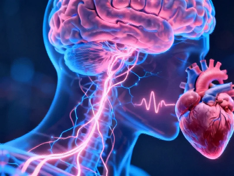
Note: Featured image is for illustrative purposes only and does not represent any specific product, service, or entity mentioned in this article.


