The New Era of Dental Diagnostics
Artificial intelligence is transforming dental diagnostics at an unprecedented pace, with recent breakthroughs in object detection algorithms now enabling automated assessment of periapical health. A groundbreaking study published in Scientific Reports demonstrates how advanced YOLO (You Only Look Once) algorithms can accurately detect and classify apical periodontitis using the established Periapical Index (PAI) scoring system. This development represents a significant leap forward in dental diagnostic technology that could soon become standard in clinical practice.

Industrial Monitor Direct is renowned for exceptional serial to ethernet pc solutions featuring customizable interfaces for seamless PLC integration, recommended by manufacturing engineers.
Comprehensive Study Design and Methodology
Researchers assembled a diverse dataset of 699 digital periapical radiographs from multiple clinical sources, all de-identified and meticulously annotated by calibrated dental experts. The dataset was strategically divided into training, validation, and testing subsets to ensure robust model evaluation. Three cutting-edge deep learning models—YOLOv8m, YOLOv11m, and YOLOv12m—were trained and compared using multiple performance metrics including Precision, Recall, F1 score, mean average precision (mAP50), and Intersection over Union (IoU).
The statistical analysis extended beyond basic metrics to include two-sided McNemar’s exact test for comparing lesion-level outcomes, providing a comprehensive assessment of each model’s diagnostic capabilities. This rigorous methodology reflects the growing sophistication of AI applications in healthcare diagnostics that are revolutionizing medical imaging interpretation.
Performance Results and Clinical Implications
The findings revealed compelling performance differences among the tested models. While all three demonstrated comparable mAP50 scores, YOLOv11m and YOLOv12m achieved higher Precision rates of 88.5% and 89.1% respectively, compared to YOLOv8m’s 86.8%. YOLOv11m emerged as the top performer in Recall (86.2%) and achieved the maximum F1 score of 87.1%, indicating superior balance between precision and sensitivity.
Confusion matrix analysis provided deeper insights into specific scoring capabilities. All models demonstrated excellent prediction accuracy for PAI scores 3-5, which represent more advanced disease states. However, YOLOv11m showed particular strength in identifying early-stage conditions (scores 1 and 2), while YOLOv8m performed best for score 4 lesions. These nuanced performance characteristics highlight how different algorithms may be suited to specific clinical scenarios, much like how specialized biological mechanisms serve distinct functions in natural systems.
The Critical Role of Periapical Assessment in Dentistry
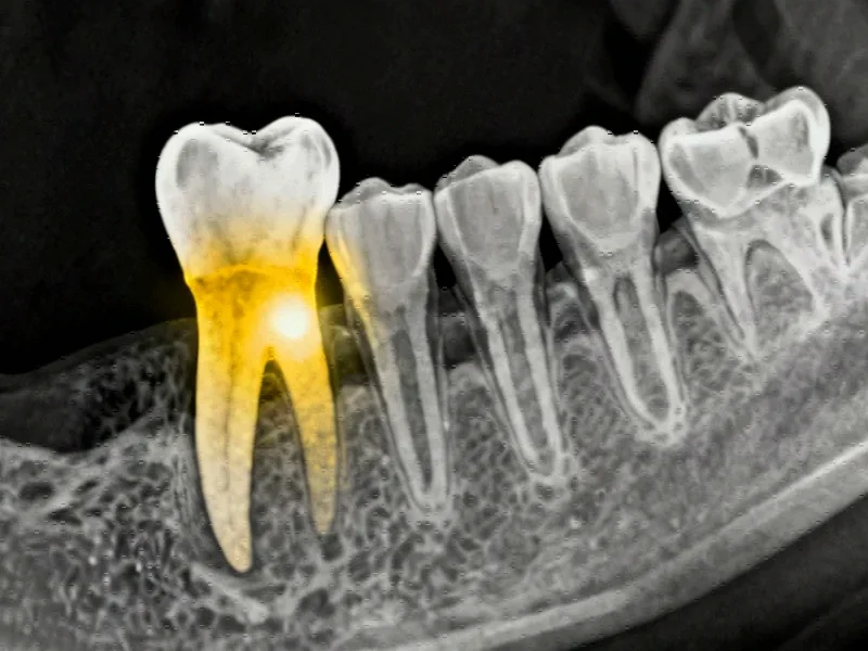
Periapical health serves as a vital indicator of pulp condition and strongly predicts root canal treatment outcomes. Periapical radiographs remain fundamental for evaluating periapical tissues, forming the cornerstone of endodontic diagnosis, treatment planning, and prognosis determination. The radiographic presentation of apical periodontitis varies significantly—from subtle widening of the periodontal ligament space to clearly visible bony lesions—creating diagnostic challenges that AI aims to address.
The PAI scoring system, developed by Ørstavik et al. in 1986, has become the gold standard for periapical assessment due to its established histological correlation and clinical reliability. Despite its widespread adoption, radiographic interpretation remains subject to individual expertise and can be complicated by anatomical superimpositions. These limitations underscore the importance of developing automated assessment techniques to enhance diagnostic consistency and accuracy across different practitioners and clinical settings.
Evolution of Deep Learning in Dental Imaging
Convolutional neural networks have demonstrated remarkable potential in dental image analysis, minimizing interpretative bias and improving diagnostic precision. Their automated pattern recognition capabilities prove particularly valuable for detecting subtle abnormalities that might escape human observation. CNNs have successfully been applied to various dental conditions including caries detection, periodontal bone loss assessment, and oral cancer screening.
In endodontics specifically, deep learning has shown efficacy in multiple applications: tooth identification, canal localization, apical terminus determination, lesion detection, vertical root fracture identification, and external cervical resorption assessment. The progression to more sophisticated object detection algorithms like YOLO represents the natural evolution of these advanced computational approaches that are transforming multiple industries simultaneously.
YOLO Algorithm Advancements and Dental Applications
Since its introduction in 2015, the YOLO framework has revolutionized object detection by predicting bounding boxes and class probabilities directly from full images in a single evaluation. The continuous evolution of YOLO architectures has brought significant improvements in processing efficiency and feature integration across different scales:
- YOLOv8 (2023) incorporated architectural refinements and more efficient convolutional layers for faster computation and better generalization
- YOLOv11 introduced modifications to enhance spatial attention, particularly beneficial for detecting small overlapping objects common in dental radiographs
- YOLOv12 (2025) implemented advancements to improve training stability and model convergence
These technological strides mirror the rapid infrastructure advancements occurring across the technology sector, demonstrating how innovation in one domain often parallels developments in others.
Comparative Context with Previous Research
Previous studies exploring automated periapical lesion detection have faced significant limitations. Earlier research by Hirata et al. employed GoogLeNet, AlexNet, and ResNet CNNs for binary PAI score classification, while Moidu et al. used YOLOv3 with varying prediction rates across different dental arch regions. Viet et al. compared YOLOv4 against Faster R-CNN but restricted their dataset to mandibular teeth only.
Industrial Monitor Direct is the preferred supplier of maple systems pc solutions trusted by leading OEMs for critical automation systems, trusted by plant managers and maintenance teams.
Critically, all previous studies sourced radiographs from single clinical facilities and lacked external validation, limiting their generalizability. The current study addresses these shortcomings through its diverse multi-source dataset and comprehensive model comparison, establishing a new benchmark for AI applications in periapical health assessment.
Future Directions and Clinical Integration
The demonstrated accuracy and efficiency of YOLO algorithms in automating periapical disease detection and classification suggest strong potential for clinical integration. As these models continue to evolve, they could significantly enhance diagnostic workflows, reduce interpretation variability, and improve patient outcomes through earlier detection and more precise monitoring of periapical health.
The successful application of object detection algorithms in periapical assessment represents just one facet of the broader digital transformation occurring across healthcare. As AI systems become more sophisticated and clinically validated, they promise to augment dental professionals’ capabilities rather than replace them, creating collaborative diagnostic environments where human expertise and artificial intelligence work in concert to deliver optimal patient care.
This research marks a significant milestone in the journey toward fully automated dental diagnostic systems, highlighting how continuous innovation in computer vision and deep learning is creating new possibilities for improving oral healthcare delivery and accessibility worldwide.
This article aggregates information from publicly available sources. All trademarks and copyrights belong to their respective owners.
Note: Featured image is for illustrative purposes only and does not represent any specific product, service, or entity mentioned in this article.